High-density MRI coil arrays with integrated field monitoring systems for human connectome mapping

High-density MRI coil arrays with integrated field monitoring systems for human connectome mapping

Magnetic Resonance in Medicine | 94 (5) 2286-2303
Purpose To develop and test two high-density MRI coil arrays with integrated field monitoring systems for enhanced diffusion imaging with strong diffusion-sensitizing gradients. Methods Two multichannel head coils were constructed for first- and second-generation 3T Connectome MRI scanners, incorporating 64 and 72 receive channels, respectively. The array coils were evaluated using RF bench-level metrics, including quality factor, tuning, matching, and coupling measurements. Imaging performance was comprehensively assessed through metrics such as SNR, 𝐵+1 efficiency, and inter-channel noise correlations, and compared with and without field camera integration. Parallel imaging capability was evaluated using geometry (g)-factors. The field camera performance was characterized by quantifying phase errors and field probe FID lifetimes. In vivo DWI acquisitions with high 𝑏-values were performed to evaluate the system's ability to correct higher-order field perturbations. Results The developed arrays demonstrated up to 1.4-fold higher SNR and superior g-factor performance when compared to a commercially available 32-channel head coil. Integration of the field camera was achieved without compromising the performance of either system. In vivo imaging with concurrent field monitoring enabled accurate spatiotemporal field corrections, significantly reducing geometric distortions, blurring, and ghosting in high 𝑏-value DWI. Conclusion The integration of high-density MRI arrays with field monitoring systems facilitated the capture and correction of spatiotemporal field perturbations during strong gradient activity, substantially enhancing image quality and diffusion parameter mapping quality. These advancements provide a robust platform for exploring the structural intricacies of the human connectome.

Purpose

To develop and test two high-density MRI coil arrays with integrated field monitoring systems for enhanced diffusion imaging with strong diffusion-sensitizing gradients.

Methods

Two multichannel head coils were constructed for first- and second-generation 3T Connectome MRI scanners, incorporating 64 and 72 receive channels, respectively. The array coils were evaluated using RF bench-level metrics, including quality factor, tuning, matching, and coupling measurements. Imaging performance was comprehensively assessed through metrics such as SNR, 𝐵+1 efficiency, and inter-channel noise correlations, and compared with and without field camera integration. Parallel imaging capability was evaluated using geometry (g)-factors. The field camera performance was characterized by quantifying phase errors and field probe FID lifetimes. In vivo DWI acquisitions with high 𝑏-values were performed to evaluate the system's ability to correct higher-order field perturbations.

Results

The developed arrays demonstrated up to 1.4-fold higher SNR and superior g-factor performance when compared to a commercially available 32-channel head coil. Integration of the field camera was achieved without compromising the performance of either system. In vivo imaging with concurrent field monitoring enabled accurate spatiotemporal field corrections, significantly reducing geometric distortions, blurring, and ghosting in high 𝑏-value DWI.

Conclusion

The integration of high-density MRI arrays with field monitoring systems facilitated the capture and correction of spatiotemporal field perturbations during strong gradient activity, substantially enhancing image quality and diffusion parameter mapping quality. These advancements provide a robust platform for exploring the structural intricacies of the human connectome.

Research Team

Prof. Dr.
Boris Keil
Principal Investigator|Scientific Coordinator
Prof. Dr.
Susanne Knake
Principal Investigator|Dep. Scientific Coordinator
Mert Murat Kurt
PhD student
Mona Alem
PhD student
Dr. med.
Felix Zahnert
Staff

Publications

  • A deep brain stimulation–conditioned RF coil for 3T MRI
    Purpose To develop and test an MRI coil assembly for imaging deep brain stimulation (DBS) at 3 T with a reduced level of local specific absorption rate of RF fields near the implant. Methods A mechanical rotatable linearly polarized birdcage transmitter outfitted with a 32-channel receive array was constructed. The coil performance and image quality were systematically evaluated using...
  • Angular dependence of detector responses in high-energy brachytherapy
    Objective To characterize three small volume ionization chambers and a diamond detector concerning their angular dependent response under Ir-192 irradiation. Approach Monte Carlo (MC) simulations as well as experimental measurements of the angular dependent response were performed at 10 cm distance in air. Further simulations under TG-43 conditions were performed at distances of 1, 3, 5 and 10...
  • Automatic detection of hippocampal sclerosis in patients with epilepsy
    Objective This study was undertaken to develop and validate an automatic, artificial intelligence-enhanced software tool for hippocampal sclerosis (HS) detection, using a variety of standard magnetic resonance imaging (MRI) protocols from different MRI scanners for routine clinical practice. Methods First, MRI scans of 36 epilepsy patients with unilateral HS and 36 control patients with epilepsy...
  • Eddy current-induced artifact correction in high b-value ex vivo human brain diffusion MRI with dynamic field monitoring
    Purpose To investigate whether spatiotemporal magnetic field monitoring can correct pronounced eddy current-induced artifacts incurred by strong diffusion-sensitizing gradients up to 300 mT/m used in high b-value diffusion-weighted (DW) EPI. Methods A dynamic field camera equipped with 16 1H NMR field probes was first used to characterize field perturbations caused by residual eddy currents...
  • High-density MRI coil arrays with integrated field monitoring systems for human connectome mapping
    Purpose To develop and test two high-density MRI coil arrays with integrated field monitoring systems for enhanced diffusion imaging with strong diffusion-sensitizing gradients. Methods Two multichannel head coils were constructed for first- and second-generation 3T Connectome MRI scanners, incorporating 64 and 72 receive channels, respectively. The array coils were evaluated using RF...

News

  • ProLOEWE mini-series "It all starts with curiosity" for World Laboratory Day
    Lisa Kempus
    23.04.2026
    To mark World Laboratory Day on 23 April, ADMIT took part in the ProLOEWE mini-series “It all starts with curiosity”. Over seven days, a daily image snippet showcased some of the research  taking place in the LOEWE laboratories and invited the community to join in the guessing game via LinkedIn. The image shared by ADMIT shows the surface receive coil, as used in magnetic resonance imaging (MRI). These...
  • First ADMIT Interdisciplinary Summer School of Medical Physics
    Lisa Kempus
    22.08.2025
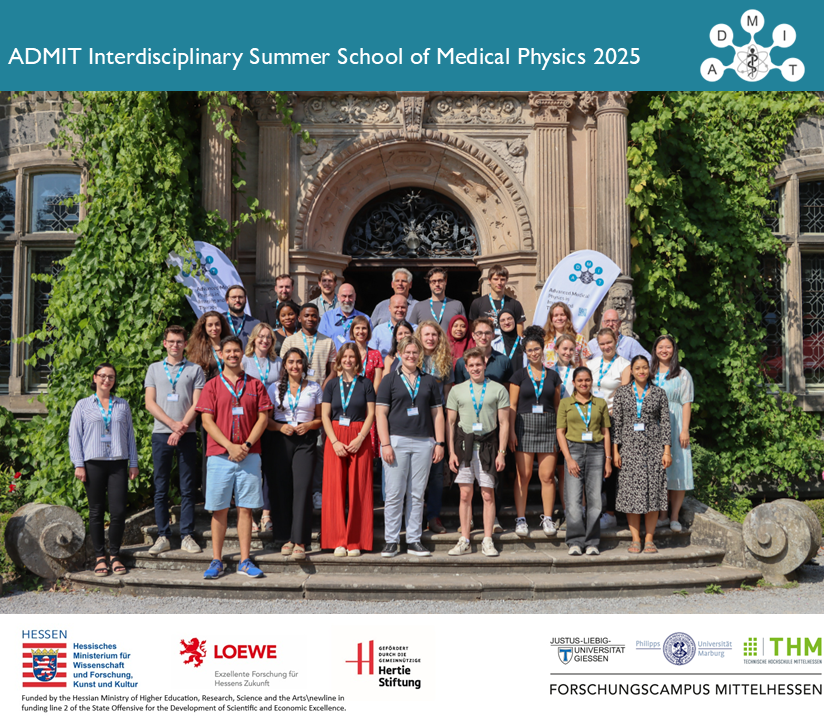
    What an inspiring and motivated group at our first ADMIT Interdisciplinary Summer School of Medical Physics, held from August 19 to 22 at Rauischholzhausen Castle!  The course brought together around 30 early-career researchers in the field of MedicalPhysics. The goal was to offer comprehensive insights into the various fields, as well as networking opportunities and interdisciplinary skills, with a special...
  • Successful end of our first ADMIT-Retreat
    ADMIT
    24.01.2025
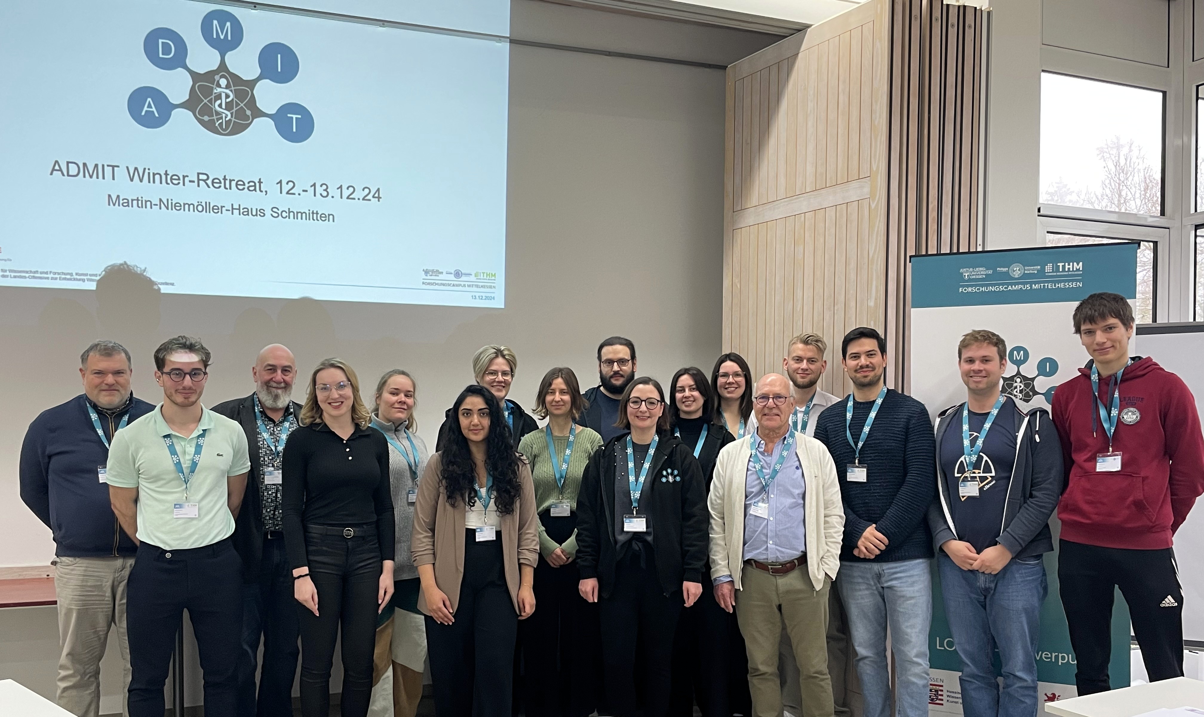
    From 12 to 13 December 2024, the members of the LOEWE Research Cluster ADMIT, in which the three Central Hessian universities of the FCMH (Justus Liebig University Giessen, Philipps University Marburg, University of Applied Sciences) have been working together since the beginning of the year, met for their first joint Retreat at the Martin-Niemöller-House in Schmitten.The focus was on an...
  • Fighting cancer and Parkinson's with medical physics
    Photo: Katrina Friese / ADMIT
    Pressestelle THM
    09.09.2024
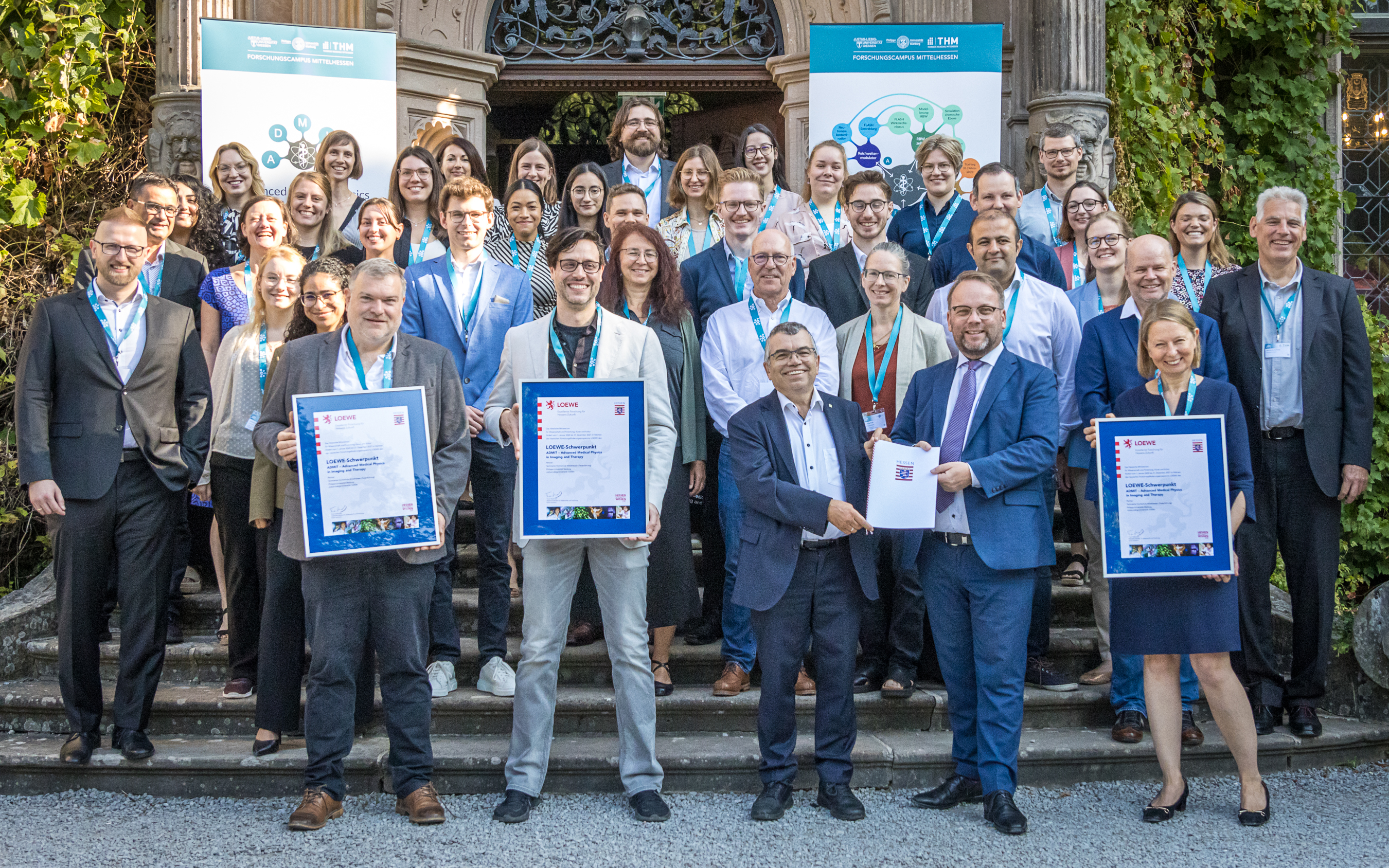
    Cancer and neurodegenerative diseases are among the greatest health and social challenges of today. Under the leadership of the THM the three universities in Central Hesse have been working together on innovative medical physics approaches in the LOEWE Research Cluster ADMIT (Advanced Medical Physics in Imaging and Therapy) since the beginning of the year in order to meet these challenges. At...
  • Boost for advances in medical technology
    Photo: THM
    Pressestelle THM
    26.03.2024
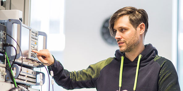
    “Brainmapping Technology”: This is the name of the LOEWE Transfer Professorship, with which the state of Hesse is supporting the utilization of Prof. Dr. Boris Keil`s research. The expert in MRI imaging procedures from the Life Science Engineering Department at THM is working on the further development of magnetic resonance imaging (MRI) for high-precision imaging of the human brain and nervous...